
FIGURE 2.
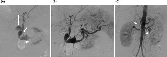
The findings and treatment of angiography. (A) Digitally subtracted image showing stenosis at the origin of the celiac trunk artery and aneurysms in the celiac artery (arrowhead), common hepatic artery (short arrow), and right hepatic artery (long arrow). However, the periphery of the right hepatic artery could not be visualized. (B) Postembolization of the right hepatic artery by multiple coils. (C) Digitally subtracted image showing multiple aneurysms (arrowheads) in both renal arteries.