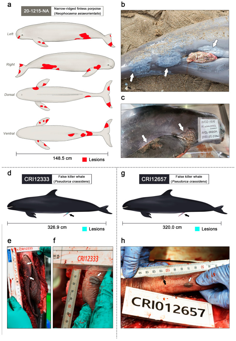
Figure 2.
Clinical lesions of gammaherpesvirus infection in the narrow-ridged finless porpoise and two false killer whales (figures were illustrated by S.B.L.). (a) The left, right, dorsal, and ventral view of the narrow-ridged finless porpoise; 20-1215-NA. Observed skin lesions are marked in red. A total of twelve skin lesions were observed on the entire body. The total body length was 148.5 cm. (b) The largest skin lesion of 20-1215-NA was 12.8 × 11.0 cm around the genital slit region (white arrows). (c) Dermatitis lesions of 20-1215-NA were found on both flippers and fluke (white arrows). (d) Genital lesions of the false killer whale CRI12333 are marked in turquoise and indicated using a black arrow. The total body length was 326.9 cm. (e) Genital lesions of the false killer whale; CRI12333 (white arrows). Numerous small vesicles were scattered on the dorsal and proximal penile epidermis and within the epidermis. The diameter of the vesicular lesions varied from approximately 1 to 4 mm. (f) The largest genital lesion of the false killer whale CRI12333 (white arrow). The pigmented vesicular lesions were located on the ventral penile epidermis and within the epidermis, and the dimensions of the lesions were 1.5 × 1.2 cm. (g) Genital lesions of the false killer whale CRI12657 are marked in turquoise and indicated using a black arrow. The total body length was 320.0 cm. (h) Small pigmented vesicular lesions with a diameter of 1–3 mm were clustered on the ventral and proximal penile epidermis and within the epidermis (black arrows). A pigmented vesicular lesion with dimensions of 1.2 × 0.7 cm was located on the left penile epidermis and within the epidermis (white arrow).

