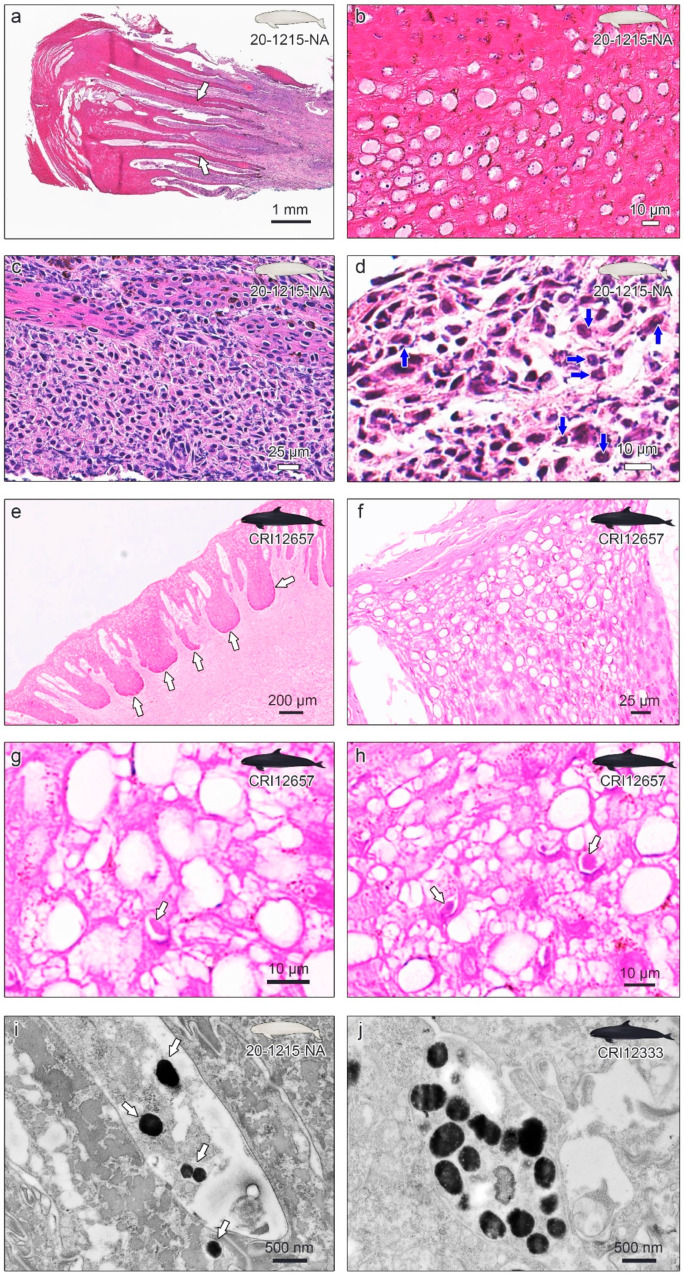
Figure 3.
Histopathological examinations of the skin lesions of narrow-ridged finless porpoise 20-1215-NA (a–d), genital lesions of false killer whale CRI12657 (e–h), and transmission electron microscopy of the 20-1215-NA and CRI12333 lesions (i,j). (a) The epidermis was moderately to markedly thickened. The white arrows indicate accentuated rete pegs into the underlying connective tissue. (b) A ballooning change is observed with amorphous eosinophilic material in the vacuoles and moderate nuclear debris within the epidermis. (c) Mononuclear cells infiltrate predominantly within the dermis at the base of the proliferative epidermis. (d) Intranuclear eosinophilic inclusion body (INI) is observed in the epidermis (blue arrows). (e) The epidermis shows the forming of thick dermal papillae rete pegs. (f) Ballooning degeneration of many nuclei is obvious. (g,h) The eosinophilic INI was observed in the epidermis (white arrows). (i) Virus-like particles from the skin lesion of 20-1215-NA were observed under the transmission electron microscope (TEM). (j) Virus-like particles from the genital lesion of CRI12333 were observed under the TEM.

