Figure 1.
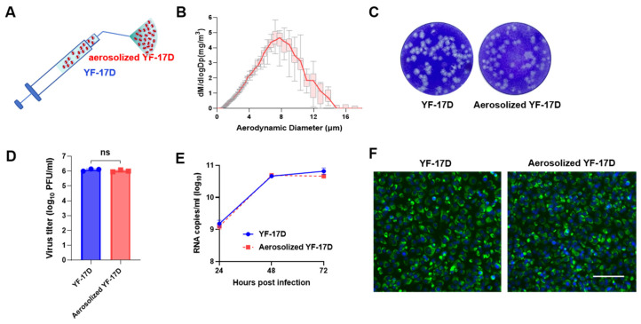
Biological characteristics of aerosolized YF-17D in vitro (A) Schematic of the hand-held liquid aerosol pulmonary delivery device (HLAPDD). (B) Aerodynamic median mass diameter (MMAD) of YF-17D aerosol measured by particle size spectrometer. (C) Plaque morphology of YF-17D and aerosolized YF-17D on BHK-21 cells. (D) The viral titers of YF-17D and aerosolized YF-17D on BHK-21 cells. (E) Replication curve of YF-17D and aerosolized YF-17D on BHK-21 cells. BHK-21 cells were infected with both liquid and aerosolized YF-17D. Cell supernatants were collected at 24, 48, and 72 hpi to measure the viral RNA load using the RT-qPCR assay. (F) Viral protein expression of YF-17D and aerosolized YF-17D on BHK-21 cells. Briefly, BHK-21 cells were infected with YF-17D and aerosolized YF-17D, then the BHK-21 cells were fixed at 48 hpi for immunofluorescence staining. The E protein of YF-17D appeared as a green fluorescence, while the cell nuclei were visualized in blue through DAPI staining. The image is referenced with a scale bar representing 100 μm. Significance was calculated using unpaired t-test (n.s., not significant).

