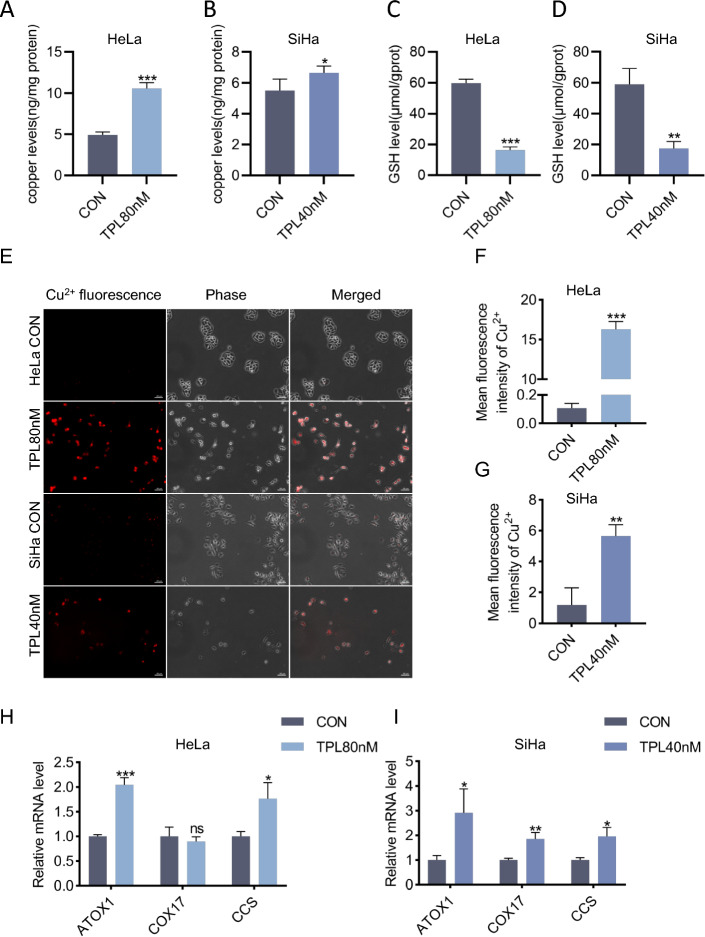
Fig. 1.
Triptolide increased the intracellular copper concentration in cervical cancer cells. A, B Copper levels were assessed by ICP‒MS in HeLa and SiHa cells treated with or without triptolide for 48 h. C, D GSH levels were assessed using a reduced glutathione assay in HeLa and SiHa cells following triptolide treatment for 48 h. E Representative images of Cu2+ fluorescence in HeLa and SiHa cells treated with or without triptolide for 48 h. Scale bars: 100 μm. F, G Mean fluorescence intensity of Cu2+ in HeLa and SiHa cells. H, I Relative mRNA levels of ATOX1, COX17 and CCS in HeLa and SiHa cells treated with or without triptolide for 48 h. (*p < 0.05, **p < 0.01, ***p < 0.001 versus the CON group.)

