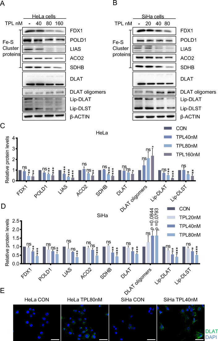
Fig. 2.
Triptolide induces cuproptosis in cervical cancer cells. A Western blotting of cuproptosis-related proteins in HeLa cells. B Western blot analysis of the levels of the apoptosis-related proteins in SiHa cells. C The protein content in HeLa cells was analyzed after triptolide treatment for 48 h. D The protein content in SiHa cells was analyzed after triptolide treatment for 48 h. E DLAT immunofluorescence after 80 nM or 40 nM triptolide treatment of HeLa and SiHa cells for 48 h (DALT-green, DAPI-blue). Scale bars: 50 μm. (*p < 0.05, **p < 0.01, ***p < 0.001 versus the CON group; ns, not significant.)

