Fig. 4.
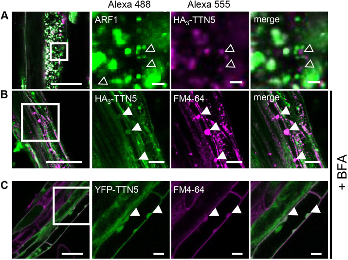
Whole-mount immunolocalization hints at TTN5 presence in BFA bodies. (A,B) Colocalization of HA3–TTN5 seedlings by whole-mount immunostaining. (A) Detection of HA3–TTN5 (anti-HA primary antibody, Alexa Fluor 555-labeled secondary antibody) with Golgi and TGN marker ARF1 (anti-ARF1 primary antibody, Alexa Fluor 488-labeled secondary antibody). Both fluorescence signals were detected in vesicle-like structures in root cells in close proximity to each other but mostly not colocalizing. The experiment was repeated twice with three seedlings (n=3). (B) Detection of HA3–TTN5 (anti-HA primary antibody, Alexa Fluor 488-labeled secondary antibody) and staining with membrane dye FM4-64 after BFA treatment (10 mM FM4-64 FX and 72 µM BFA for 1 h). Alexa Fluor 488 signals colocalized with FM4-64 in BFA bodies in root cells. The experiment was repeated three times with three seedlings (n=3). (C) YFP fluorescence in YFP–TTN5 seedlings, co-analyzed with FM4-64 after BFA treatment. YFP fluorescence signals colocalized with FM4-64 in BFA bodies similar to what was seen in B. The experiment was performed once with three independent YFP–TTN5 lines (n=3). Colocalization indicated by filled white arrowheads, non-colocalized HA3–TTN5 Alexa Fluor 488-labeled signals is indicated with empty white arrowheads. Scale bars: 50 µm (overview images on left), 10 µm (magnifications).

