Fig. 6.
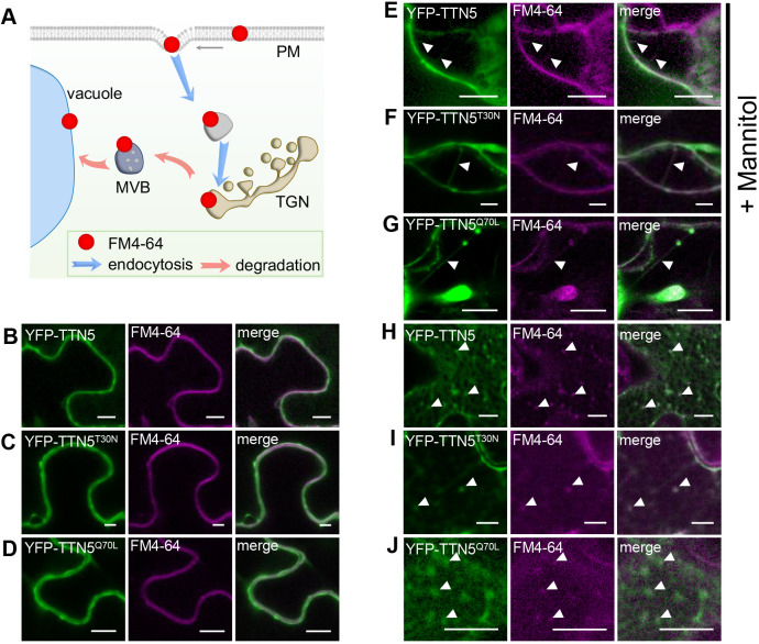
TTN5 might colocalize with endocytosed PM material. (A) Schematic representation of the progressive stages of lipophilic membrane dye FM4-64 localization and internalization in cells. After infiltration, it first localizes in the PM, and later in intracellular vesicles and membrane compartments, reflecting the endocytosis process (Bolte et al., 2004). (B–J) YFP fluorescence colocalized with FM4-64 in N. benthamiana leaf epidermal cells as observed by confocal microscopy, following transient expression of YFP–TTN5, YFP–TTN5T30N and YFP–TTN5Q70L. (B–D) YFP signals colocalized with FM4-64 at the PM. (E–G) PM localization of YFP fluorescence was evaluated after mannitol-induced plasmolysis (1 M for ∼15–30 min). Formation of Hechtian strands is a sign of PM material and fluorescence staining there (filled arrowheads). (H–J) Internalized FM4-64 was present in vesicle-like structures that showed YFP signals. Colocalization indicated with filled arrowheads. Experiments were repeated three times with two plants (n=2). Scale bars: 10 μm.

