. 2024 Aug 13;25(9):807–823. doi: 10.3348/kjr.2023.1307

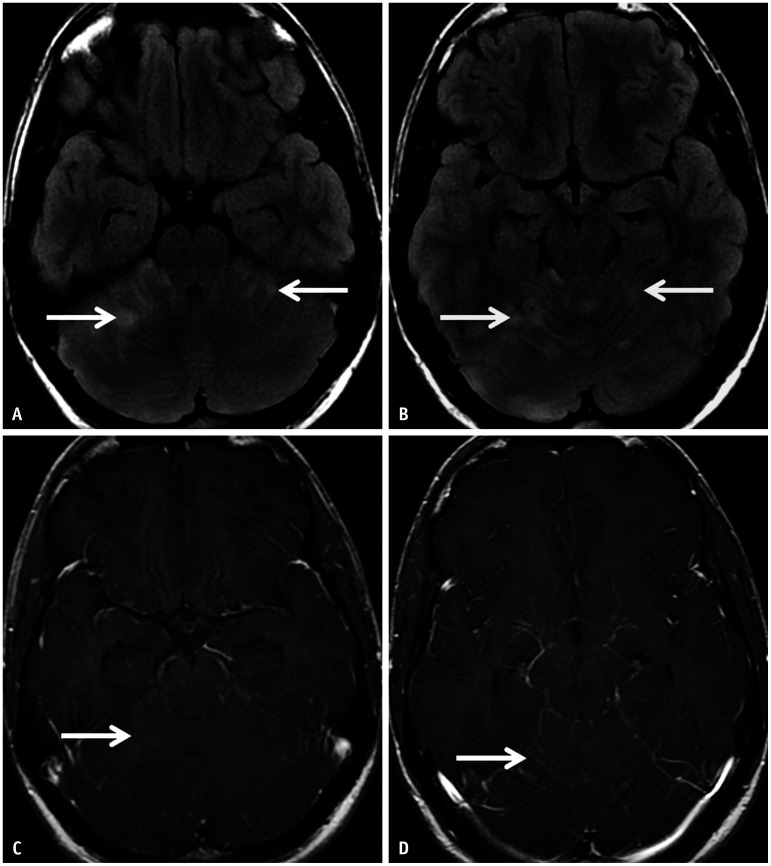
Fig. 8. Autoimmune cerebellitis in a 19-year-old patient with anti-VGKC encephalitis presenting with a one-month history of arm twitching (right > left). A, B: Axial FLAIR images reveal patchy, ill-defined hyperintensities involving the cerebellar hemispheres (arrows), more on the right. C, D: Contrast-enhanced-T1 images reveal questionable faint enhancement in the right cerebellar lesions (arrow in C) and subtle leptomeningeal enhancement along the right interfolial spaces (arrow in D). Anti-LGI1 encephalitis (a subgroup of anti-VGKC encephalitis) is known to present with cerebellar involvement. VGKC = voltage gated potassium channel.

Fig. 8