. 2024 Mar 28;5(1):3–20. doi: 10.12336/biomatertransl.2024.01.002

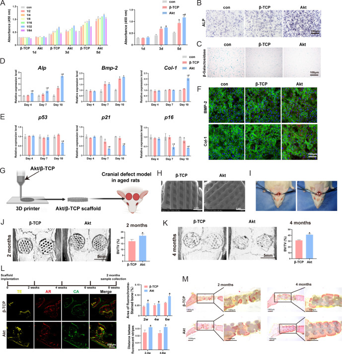
Figure 3. Akt promote senescent bone regeneration in vitro and in vivo. (A–F) The effects of Akt onproliferation (A), ALP staining (B), β-galactosidase staining (C), osteogenic-related genes (D), senescent-related genes (E) and osteogenic-related proteins (F) in O-BMSCs. (G) The schematic diagram of 3D printed Akt/β-TCP scaffold. (H) The SEM images of Akt/β-TCP scaffold. (I) Critical skull bone defects model of senescent rats. (J–M) The bone repair evaluation of 3D printed Akt/β-TCP scaffold by micro-CT, sequential fluorescence and VG staining. Data are expreesed as mean ± SD. *P < 0.05, vs. β-TCP; #P < 0.05, vs. α-MEM without extracts. Scale bars: 100 μm (B, C, F, L), 1 mm (H), 5 mm (J, K). Reprinted from Qi et al.61 3D: three-dimensional; Akt: akermanite; Alp: alkaline phosphatase; AR: alizarin red; Bmp-2: bone morphogenetic protein 2; BV/TV: bone volume fraction; CA: calcein; Col-1: type I collagen; CT: computed tomography; O-BMSCs: aged bone marrow mesenchymal stem cells; SEM: scanning electron microscopy; TE: tetracycline; VG: Van Gieson; α-MEM: α-minimal essential medium; β-TCP: β-tricalcium phosphate.

Figure 3.