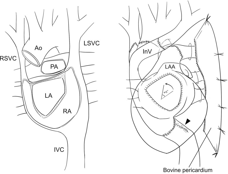
Fig. 2.
Schematic images after cardiectomy (left) and heart transplantation (right): the left atria were anastomosed with a 45-degree counterclockwise rotation of the donor heart. The recipient IVC was lengthened by a conduit made of residual RA tissues (arrowhead). A bovine pericardium was used to create a screen for excluding the left lung. Ao aorta, InV innominate vein, IVC inferior vena cava, LA anatomical left atrium, LAA left atrial appendage, LSVC left superior vena cava, PA pulmonary artery, RA anatomical right atrium, RSVC right superior vena cava

