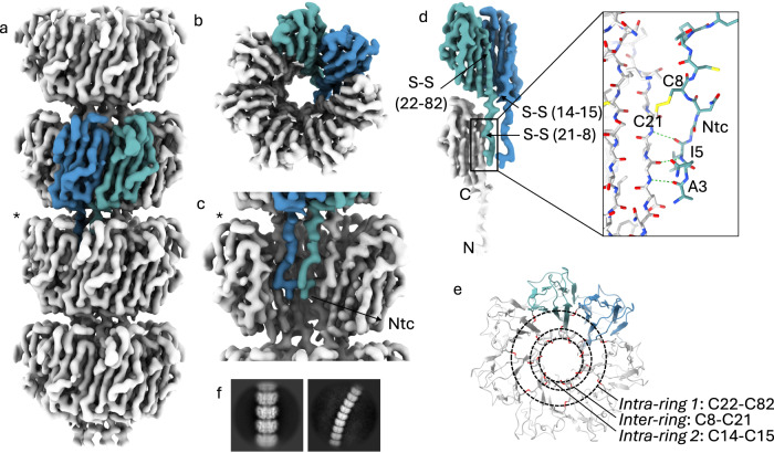
Fig. 2. Cryo-EM volume of recEna3A L-ENA fibers.
a Helical ultrastructure of L-ENA determined at 3.32 Å global resolution (cut-off criterion at FSC = 0.143 Å−1) revealing an axial stacking of heptameric Ena3A rings that rotate 18.5° clockwise relative to each other. Two neighboring Ena3A monomers are colored in blue and cyan, b top-view of a single ring with a highlight of the β-sheet augmentation between the blue and cyan subunit, c zoom-in of a single ring - for clarity, two subunits were removed from the ring highlighted with an asterisk in (a) - showing the docking of the N-terminal connectors that covalently tether the ring docked above, d Highlight of the lateral intra-ring contacts: two types of disulfide bridges (Cys14-Cys15; Cys22-Cys82) exist within two neighboring subunits. In turn, each subunit connects to a subunit in the ring below via their Ntc (Cys8-Cys21; inset in stick representation), e On-axis top-view of the L-ENA fiber model in cartoon with disulfide bridges shown in red; f 2D class averages of straight and curved L-ENA segments covering 4 and 9 rings, respectively.

