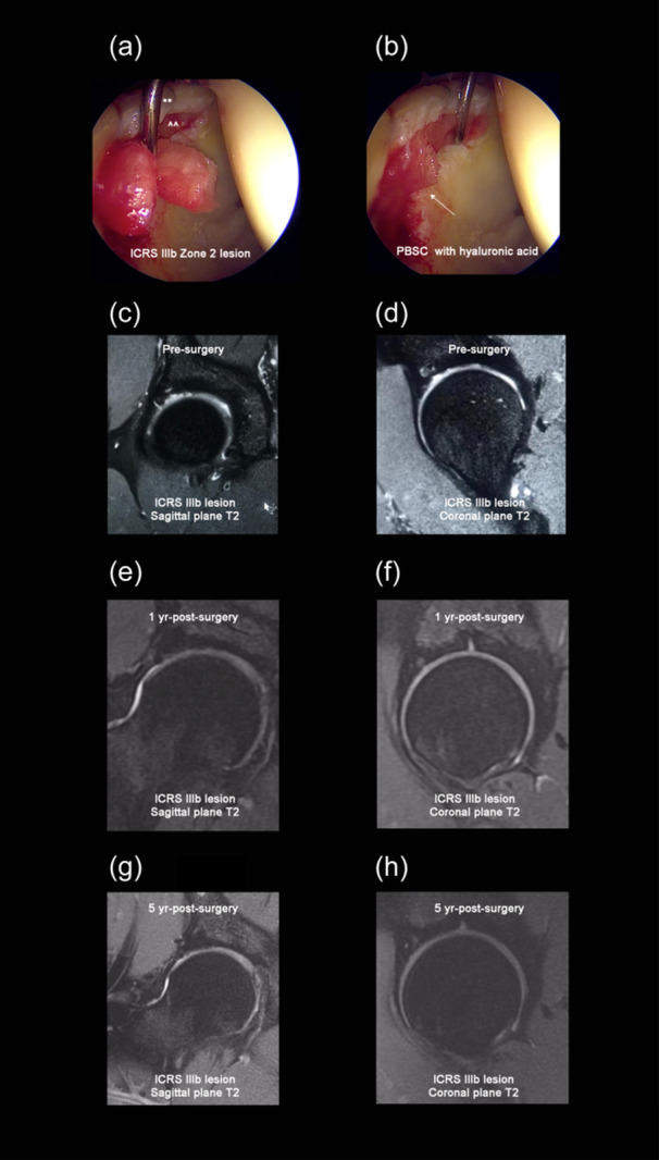
Figure 1.

(a) 34‐year‐old male with International Cartilage Repair Society (ICRS) IIIb Zone 2 osteochondral lesion during the placement of the infiltration of peripheral blood stem cells (PBSC) with hyaluronic acid. ** = repaired labrum. ^^ = chondral lesion. (b) Final placement of the PBSC with hyaluronic acid (white arrow) in the chondral lesion for the same 34‐year‐old male patient. (c and d) A T2 sagittal and coronal MRI of a 24‐year‐old male with ICRS IIIb lesion. (e and f) A T2 sagittal and coronal MRI of the same 24‐year‐old male patient at 1 year post‐surgery. (g and h) A T2 sagittal and coronal MRI of the same 24‐year‐old male patient at 5 years postsurgery showed a complete cartilage lesion repaired, stable, and complete subchondral bone repaired.
