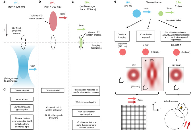
Fig. 1. Rationale for a preferred two-photon fluorophore photoactivation in the visible wavelength range, and nanoscopic imaging modalities.
Optical sectioning is achieved by the confocal detection principle (use of a pinhole), in addition to selective activation of photoactivatable fluorophores in targeted sample volumes. Only activation at the visible (green) wavelength allows matching of the rendered fluorescence to the detection volume in z (dashed lines), because the focusing performance is highly corrected for visible light and especially in the green range. Ultraviolet (UV) and near-infrared (NIR) light is focused with substantial chromatic offsets. a One-photon activation (1PA) with UV light leads to continuous activation along the beam path. b For 2PA of fluorophores, shown here for the case of near-infrared light, the activated volume is sharply confined as the volume with sufficiently high photon density for ensuing the 2-photon activation (2PA) process. This leads to background suppression outside. c 2PA with visible light, the concept explored in this work for sectioning in fluorescence nanoscopy with 515-nm femtosecond pulses. d Features (advantages and disadvantages) of the visible-light 2PA vs. UV 1PA approach. e Examples of imaging modes demonstrated in conjunction with 515-nm 2PA. STED (stimulated emission depletion), MINSTED: nanoscopy concepts.

