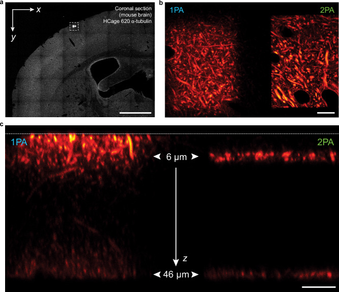
Fig. 5. Two-photon photoactivation with 515 nm light provides selective access to optical sections in mouse brain tissue.
To compare 1PA at 375 nm and 2PA at 515 nm, two layers at z = 6 µm and z = 46 µm were activated within two adjacent rectangular regions in a mouse brain cross section with HCage 620 labeling α-tubulin. a Confocal overview image after activation of two areas with 1PA (left) and 2PA (right). b Enlarged view of region shown boxed in (a), with the two activated regions. c A cross-sectional y-z scan shows the activated layers 6 µm and 46 µm inside the tissue slice (1PA on the left vs. 2PA on the right). The white dashed line at the top indicates the location of the coverglass. Two distinct layers are clearly visible in the 2PA case. Scale bars: 1 mm a, 10 µm b, c.

