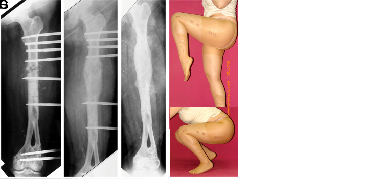
Figure 3.
(A) MRI image of an osteosarcoma located in the distal femur of a 7-year-old girl (initial sarcoma surgery performed by Professor Harzem Ozger). (B) The patient underwent a double barrel free vascularized fibula graft. An orthoroentgenogram taken 11 years later shows an 8 cm shortening in the patient. (C-G) Serial roentgenograms taken after lengthening from the femoral diaphysis with a unilateral monorail fixator and the postoperative functional outcome.

 Content of this journal is licensed under a