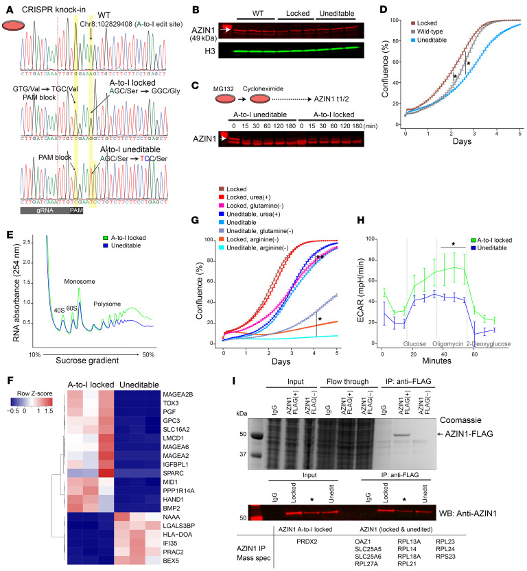
Figure 3. Azin1 A-to-I–uneditable state hinders cell growth and limits glycolytic capacity.
(A) Sanger sequencing chromatograms for wild-type (HEK293T; top), AZIN1 A-to-I–locked (middle), and AZIN1 A-to-I–uneditable homozygous cell lines (bottom). Homology-directed repair donor oligonucleotides used for CRISPR knockin are shown in Supplemental Figure 5A. (B) Western blotting for AZIN1 under indicated conditions (~70% confluence). (C) Determination of AZIN1 protein turnover under indicated conditions. Nascent protein synthesis was inhibited with 250 μg/mL cycloheximide. Arrow points to AZIN1. Bands below AZIN1 result from inhibition of proteasomal degradation with MG132. n = 2 biological replicates. (D) Real-time monitoring of cell growth for AZIN1 A-to-I–locked, uneditable, and wild-type cells. n = 3 independent experiments with n = 6 technical replicates for each experiment. *P < 0.05 at all time points for indicated conditions, except the stationary phase between AZIN1 A-to-I–locked and wild-type cells. Representative images are shown in Supplemental Figure 5C. (E) Polyribosome profiling of AZIN1 A-to-I–locked and uneditable cell lines. n = 3 independent experiments. Mean polysome/monosome ratios for A-to-I–locked and uneditable genotypes are 4.1 and 3.6, respectively. (F) Heatmap of the top 20 differentially expressed genes between AZIN1 A-to-I–locked and uneditable cell lines as determined by RNA-Seq (https://connect.posit.iu.edu/azin1/). (G) Cell growth under indicated conditions. Representative images are shown in Supplemental Figure 5D. *P < 0.05, **P < 0.05 after day 1 and day 2.5 for indicated conditions, respectively. (H) Extracellular acidification rates under indicated conditions (Seahorse glycolysis stress test). n = 3 independent experiments with n = 3 technical replicates for each experiment. *P < 0.05 vs. AZIN1-uneditable cells at indicated time points. (I) Identification of AZIN1-interacting molecules by mass spectrometry. Top: Coomassie staining for input, flow-through, and immunoprecipitated unfractionated lysates from IgG control and transfection of FLAG-tagged AZIN1 or AZIN1 without FLAG plasmids. Middle: Western blotting for AZIN1. Cells overexpressing FLAG-tagged A-to-I–locked AZIN1 or uneditable plasmids were fractionated into cytoplasmic and nuclear compartments and immunoprecipitated using anti-FLAG antibody (cytoplasmic fraction is shown). See also Supplemental Figure 5H. Summary of coprecipitated proteins with AZIN1 is presented in the bottom table. n = 3 independent experiments. *Plasmid construct not used in this article.

