Fig. 7.
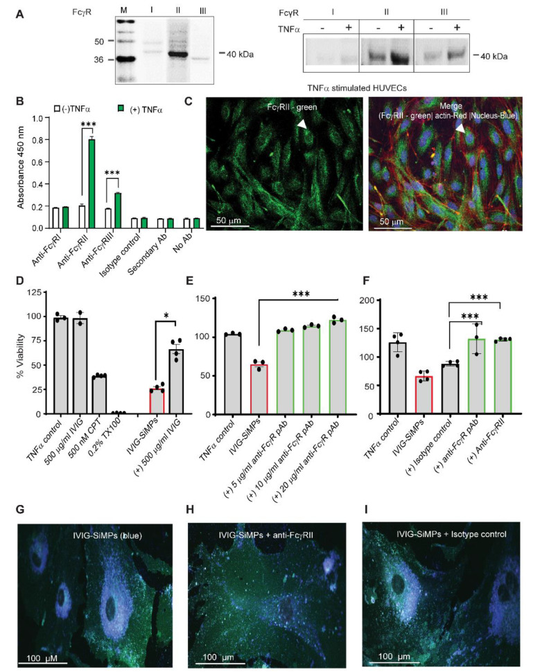
Cellular uptake and toxicity of 200 nm IVIG-SiMPs in TNFα-stimulated HUVECs is FcγRII dependent. FcγRII expression is increased in TNFα-stimulated HUVECs. The FcγR expression was evaluated by (A) Western blot, (B) ELISA, and (C) immunofluorescence (IF) cytostaining using FcγR specific antibodies. For IF staining, FITC conjugated FcγRII was utilized, along with nuclear staining (Hoechst 33342-blue), and actin tracking staining CellMask Deep Red. The slides were mounted using gold antifade mounting media (Invitrogen) and imaged using a confocal microscope (LSM700) with a 40x oil objective. Scale bars represent 50 μm. Arrows show FcγRII expressed in TNFα-stimulated HUVECs. (D-F) Soluble IVIG and anti-FcγR antibody markedly decreased the toxicity of 200 nm IVIG-SiMPs (100 µg/ml; 24 h.) in TNFα-stimulated HUVECs. TNFα -stimulated HUVECs were pre-incubated with (D) 500 µg/ml IVIG or (E) anti-FcγR pAb, or (F) anti-FcγRII mAb for 2 h. at 37 oC. The cells were then incubated with IVIG-SiMPs for an additional 24 h. The percentage viability of HUVECs was measured by CCK-8 assay. The results are representative data of at least three independent experiments, *p < 0.05. (F) Pre-incubation of TNFα-stimulated HUVECs with anti-FcγRII mAb decreased the uptake of 200 nm IVIG-coated blue, fluorescent SiMPs assessed by LSCM. HUVECs were incubated with 200 nm IVIG-coated blue, fluorescent SiMPs (100 µg/ml) (G) without or pre-exposure with (H) 10 µg/ml anti-FcγgRII mAb or (I) the corresponding isotype control mAb. Then the cells were stained with cell mask green for 30 min at 37 oC and subjected to LSCM analysis. Mean ± SEM, n = 3, * p < 0.05, ** p < 0.01, *** p < 0.001

