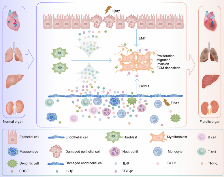
Fig. 1.
Schematic diagram of the mechanism of organ fibrosis. When tissues and organs such as the heart, liver, lung and kidney are subjected to sustained external injury, damaged parenchymal cells initiate injury-related molecular patterns, secrete inflammatory mediators, recruit inflammatory cells, and promote inflammation in damaged tissues. Quiescent fibroblasts in these organs are activated to become myofibroblasts in response to the body's repair response to damaged tissues, and epithelial-mesenchymal transition (EMT) and endothelial-mesenchymal transition (EndMT) further increase the number of myofibroblasts in damaged organs. Myofibroblasts continuously secrete extracellular matrix (ECM), such as α-smooth muscle actin (α-SMA), collagen I (COL I), and fibronectin (FN), which leads to the development of organ fibrosis [7, 67–70]

