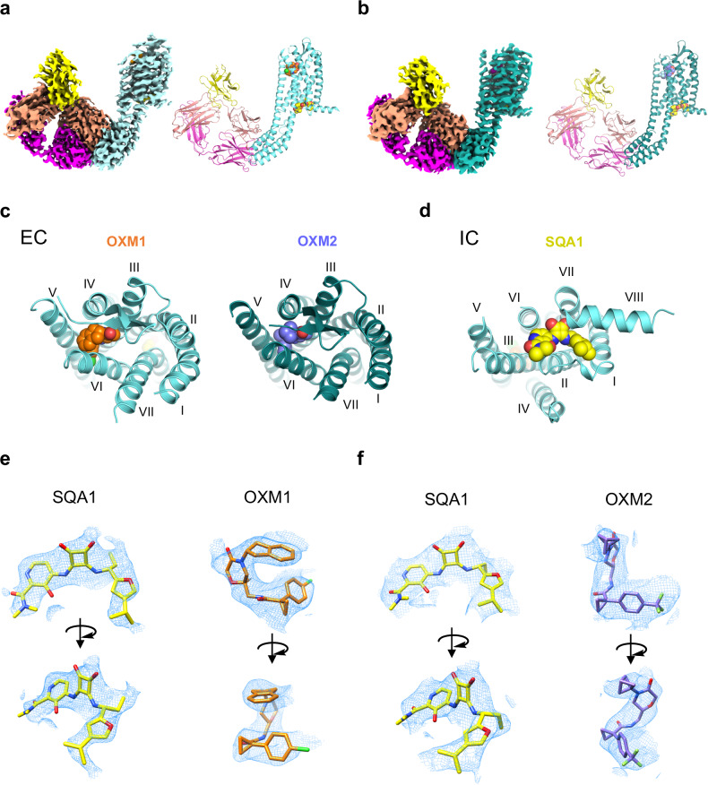
Fig. 2. Overall structures of CCR6 in complex with OXM and SQA analogues.
a,b Cryo-EM map (left) and model (right) of CCR6 complex with anti-BRIL Fab and anti-Fab Nb bound to a OXM1 (orange carbon spheres) and SQA1 (yellow carbon spheres), or b OXM2 (slate carbon spheres) and SQA1 (yellow carbon spheres). Protein is coloured by subunit as follows: Fab heavy chain, magenta; Fab light chain, salmon; Nb, yellow; CCR6, aquamarine in a and deep teal in b. c Extracellular (EC) view of CCR6 bound to OXM analogues, OXM1 (left) and OXM2 (right). d Intracellular (IC) view of CCR6 bound to the SQA analogue, SQA1. Both c and d use the same colour code as a and b. e SQA1, OXM1, and f SQA1, OXM2 fits into the cryo-EM density maps of the two structures from two viewing angles.

