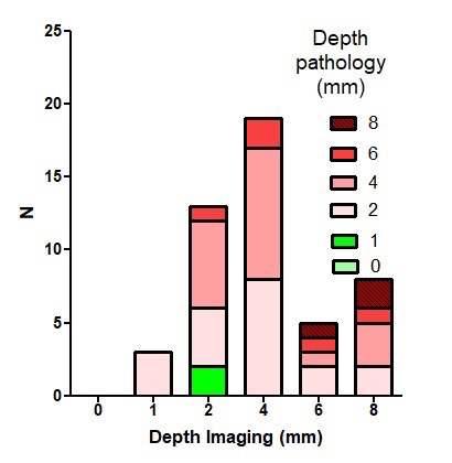
Figure 4.

Diagnosis of isthmocele based on depth measurements by imaging and pathology (macroscopically): Imaging measurements (mm) illustrated with a frequency distribution (bars) and in each of those bars, the pathology measurements (mm) illustrated by different colors. Using the isthmocele definition as depth ≥ 2mm; imaging missed 3 isthmoceles (first column from left that showed 1mm by imaging while the color is pink (2mm by pathology)) and diagnosed 2 isthmoceles (second column from left that showed 2mm by imaging with green color representing 1mm by pathology). This also explains that isthmocele prevalence at our series was 94% by imaging and 95% by pathology (macroscopically). Equally important is to realise that deep isthmoceles of 8mm by imaging (last column from right) vary between 2 and 8 mm by pathology (represented by colors at that column). Suggesting repeat imaging measurements for confirmation before deciding on surgery.
