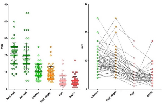
Figure 7.

Histology (macroscopic) measurements of anterior uterine wall thickness (18 mm,10.5-30), posterior uterine wall thickness (22 mm, 13.5-26.5) and isthmus or adjacent myometrial thickness (AMT) (10 mm, 6-15). The sum of RMT and depth of the isthmocele (RMT+ depth) is similar to the AMT (Isthmus), thus indirectly confirming the accuracy of those measurements. The right graph shows the individual measurements in relation to each other. Occasional variations can be noticed. It also showed that deeper isthmoceles (red dots) are associated with thinner RMTs (pink dots).
