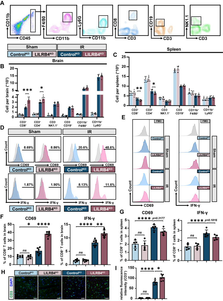
Fig. 6.
LILRB4 deficiency in microglia aggravates cytotoxic CD8+ T cells accumulation in brain after cerebral ischemia. (A) Flow-gated strategy after isolation of immune cells from brain and spleen 1 day after tMCAO in Control and LILRB4-KO mice. Fluorescent negative control was used. (B) Calculation of macrophages, neutrophil, CD4+ T cells, CD8+ T cells, NK cells, and B cells in the brains of Control, LILRB4-KO mice 1 day after tMCAO. The data show the absolute value of each brain. (n = 8; ***p = 0.0002, *p = 0.0102). (C) Quantification of macrophages, neutrophil, CD4+ T cells, CD8+ T cells, NK cells, and B cells in the spleens of Control, LILRB4-KO mice 1 day after tMCAO. The data show the absolute value of each spleen. (n = 8; **p = 0.0033, *p = 0.0192). (D–G) The expression of CD69 and IFN-γ on CD8+ T cells in the brain (D and F) and spleen (E and G) by FACS analysis for 1 day after tMCAO. (n = 5/6; *p = 0.0429, **p = 0.0017, ****p < 0.0001). (H) Representative immunofluorescence staining plots of T lymphocyte (anti-CD3, green; DAPI staining for nuclei, blue) at the infarct border site in Control, LILRB4-KO mice 1 day after tMCAO. Scale bar, 200 μm. (I) Quantification of data in (H). (n = 7; *p = 0.0376, ****p < 0.0001)

