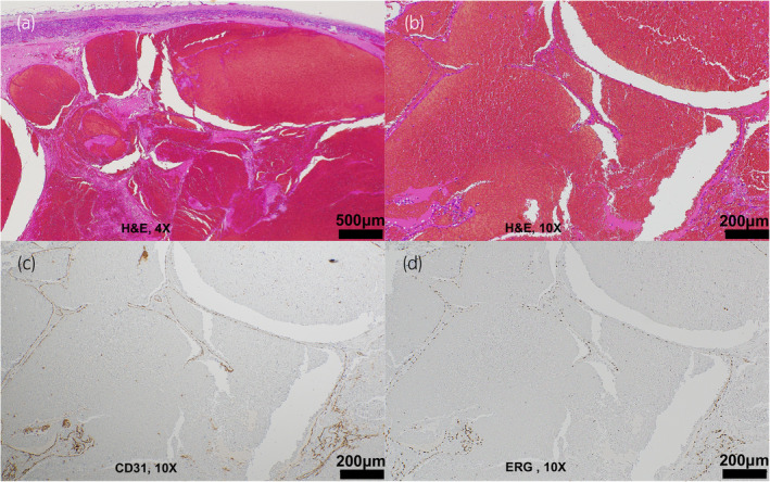
Fig. 2.

Pathological findings of the tumor. (a, b) Thin‐walled cavernous blood vessels are observed at the margin of the tumor. (c) Immunostaining confirmed CD31 positivity in the cytoplasm of the vascular endothelium. (d) Immunostaining confirmed ERG positivity in the nuclei of the vascular endothelium.
