. 2024 Aug 30;12(8):e009160. doi: 10.1136/jitc-2024-009160

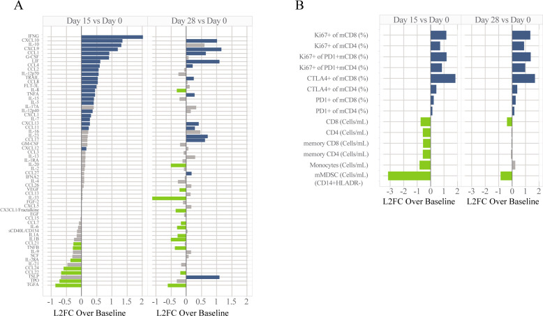
Figure 1. Peripheral blood analyses for MARIO-3. Analytes for cytokine (A) and immune cell subsets (B) determined by Luminex and flow cytometry, respectively are expressed as log2FC over baseline for samples collected at day 15 and day 28 after treatment. Absolute cell counts for monocytic MDSCs (HLAR-CD14+CD11b+CD33+), monocytes (HLADR+CD14+CD11b+CD33b+), CD8+T cells, CD4+T cells, memory CD8 T-cells (mCD8; CD45RA-CD8+CD3+), memory CD4 T cells (mCD4; CD45RA-CD4+CD3+) and subpopulations including per cent of memory CD8/CD4 T-cells expressing exhaustion markers PD-1 and CTLA-4, proliferation (Ki67+) of memory CD8/CD4 T-Cells, and proliferation (Ki67+) of exhausted (PD1+) memory CD8/CD4 T-Cells are shown (B). One-way repeated measures ANOVA was performed with colors representing significance and direction of change; blue: significant upregulation, green: significant downregulation, gray: not significant. For cytokine analysis, p-values were adjusted using Bonferroni corrections for multiple comparisons. ANOVA, analysis of variance; CTLA-4, cytotoxic T-lymphocytes-associated protein 4; MARIO-3, MAcrophage Reprogramming in Immuno-Oncology-3; MDSCs, myeloid-derived suppressor cells; PD-1, programmed cell death protein-1.

Figure 1