. 2024 Aug 28;12(8):e009869. doi: 10.1136/jitc-2024-009869

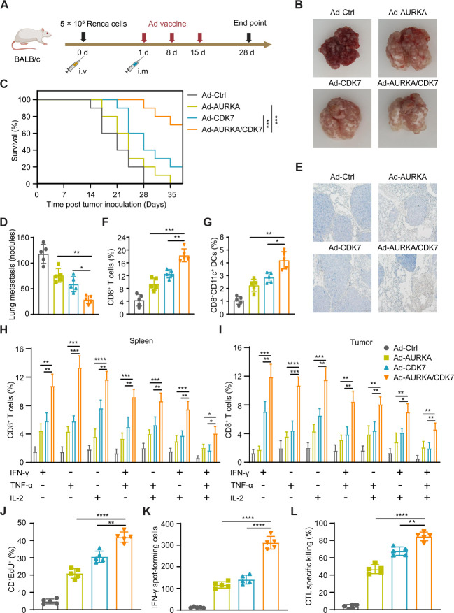
Figure 6. Ad-AURKA/CDK7 treatment suppressed tumor metastases by activating multifunctional CD8+ T cells. (A) A schematic diagram displayed the design and treatment of lung metastasis. (B) The representative image of lung metastasis nodules in each group. (C) The survival curve of mice in different groups after Renca tumor lung metastasis (n=10 mice per group). (D) The number of metastatic nodules was counted on the lung surface of immunized mice (n=5 mice per group). (E) Typical immunohistochemistry image of the lung-infiltrating CD8+ T cells in each group immunized with different vaccines. The scale was 200 µm. (F, G) The proportion of CD8+ T cells and CD8+CD11c+ DCs in the lungs of treated mice in each group. (H, I) Percentages of multifunctional CD8+ T lymphocytes producing IFN-γ, TNF-α, or IL-2 in spleens and tumor tissues of different groups. (J) The EdU assay was used to detect the proliferation of CD8+ T cells. (K) The number of IFN-γ-secreting T lymphocytes was counted using ELISpot assay. (L) The percentages of tumor-specific killing capability of CTL in each group. One-way analysis of variance was used for comparisons among multiple groups. Survival analysis was performed using the log-rank (Mantel-Cox) test. The data expressed as means±SD. *p<0.05, **p<0.01, ***p<0.001, and ****p<0.0001. Ad, adenovirus; AURKA, Aurora kinase A; CDK7, cyclin-dependent kinase 7; CTL, cytotoxic T lymphocyte; DC, dendritic cell; ELISpot, enzyme-linked immunosorbent spot; IFN, interferon; IL, interleukin; i.m, intramuscular; i.v, intravenous; TNF, tumor necrosis factor.

Figure 6