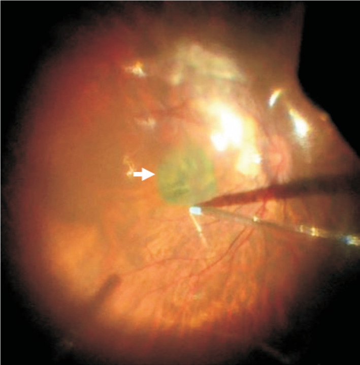
Figure 2. Covering the CSL on the MH.

A piece of 3×3 mm2 pre-processed CSL (white arrow) was passed the vitreous cavity through a 25-gauge valved trocar and laid flat on the surface of MH. The position of CSL should be in the middle of the MH, and the CSL should cover the edge of the MH. The residual liquid around the CSL was removed with a flute needle to keep the CSL tightly attaching retina and no longer moving. CSL: Corneal stromal lenticule; MH: Macular hole.
