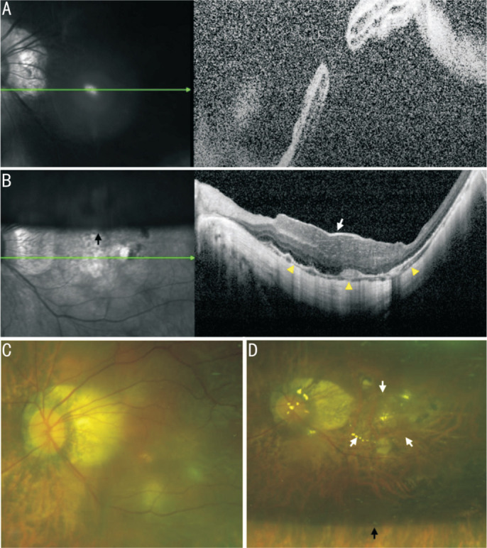
Figure 3. Information of the No.4 patient.

A, C: The OCT and SLO of macular area show an MHRD before PPV; B, D: The OCT and SLO of macular area show that RD was reattached and MH was healed by covering CSL (white arrow) after PPV. The yellow triangles point to ellipsoid zone of continuity healing. The black arrow points to the boundary of the C3F8 gas that had not yet been absorbed. OCT: Optical coherence tomography; SLO: Scanning laser ophthalmoscopy; PPV: Pars plana vitrectomy; RD: Retinal detachment; MH: Macular hole; MHRD: Macular hole retinal detachment; CSL: Corneal stromal lenticule.
