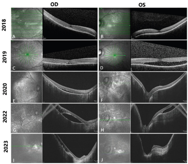
Figure 2. Consecutive optical coherence tomography images from 2018 to 2023.
Horizontal scan line length: 9.0 mm (A, B); 6.0 mm (C, D); 12 mm (E to H); 16 mm (I, J). In 2018, shallow retinal detachment with patchy subretinal hyperreflective foci and thinning of the outer nuclear layer (ONL) was noted in the right eye (RE, A), while the left eye (LE) showed thinning of ONL and intensively hyperreflective retinal pigment epithelium (RPE, B). In 2019, the subretinal fluid increased and the retina was thinned out in both eyes (C, D). In 2020, intraretinal cystic spaces appeared in the RE (E), and the subretinal hyperreflective materials was noted in the LE (F). In 2022, the outer plexiform layer was absent, intraretinal cystic spaces increased and the excavation of retinal tomography deepened in both eyes (G, H). At the last visit (2023), the excavation of retinal tomography further deepened, choroidal hyperreflectivity intensified, and the retina became much thinner in both eyes (I, J).

