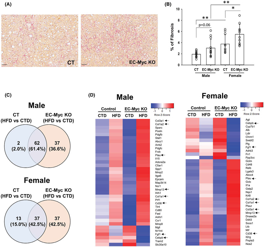
FIGURE 6.
Endothelial c-Myc deletion exacerbates HFD-induced liver fibrosis. (A) Representative images of picrosirius red-stained liver sections of CT and EC-Myc KO livers after long-term exposure to HFD. Scale bar, 50 μm. (B) Quantification of fibrosis. Results represent the percentage of positively stained area relative to total tissue area (n = 8–13). (C) Venn diagrams indicating the number of fibrosis-related genes in male (top panel) and female (bottom panel) mice significantly altered in control and EC-Myc KO livers after short-term exposure to HFD (n = 3–4). (D) Heatmaps showing fibrosis-related genes significantly altered in male (left panel) and female (right panel) mice exclusively in EC-Myc KO livers after short-term exposure to HFD (n = 3–4). Genes marked with black arrows are present in both male and female animals. CT, control; CTD, low-fat control diet; EC-Myc KO, endothelial c-Myc knockout; HFD, high-fat diet. *p < .05, **p < .01

