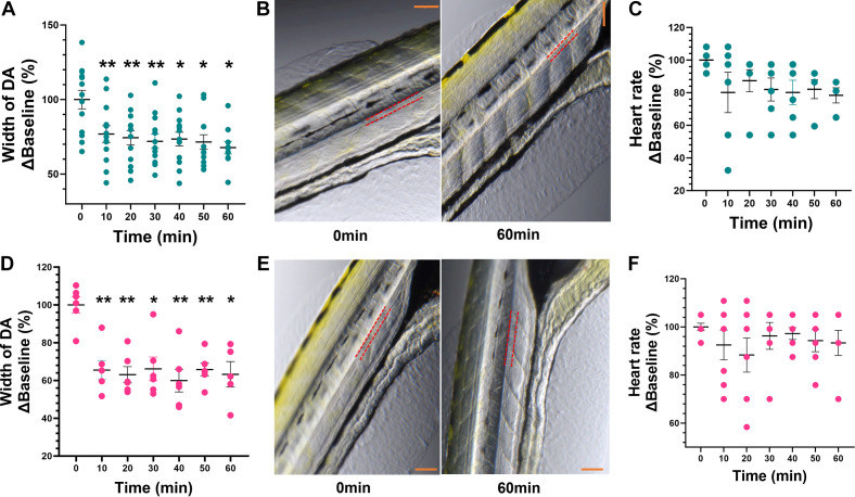
Figure 4.
Effects of phenylephrine and angiotensin II (ANG II) on vascular tone and heart rate in anesthetized zebrafish larvae. A and D: administration of 1 µM ANG II (A) and 100 µM phenylephrine (PE) (D) produced a decrease in the cross-sectional width of dorsal aorta (DA) at all time points when compared with baseline (0 min). B and E: representative images at baseline (0 min) and the 60-min timepoint for ANG II (top) and PE (bottom), with red dotted lines marking the outline of the DA. Scale bar in orange = 200 µm. C and F: ANG II (C) and (F) PE had no effect on heart rate (beats/min). Data are presented as means ± SE (n = 6–7 PE and 6–13 ANG II) and calculated as %Baseline (0 min). Repeated-measures one-way ANOVA with a Dunnett’s post hoc test; *P < 0.05, **P < 0.01.

