FIGURE 2.
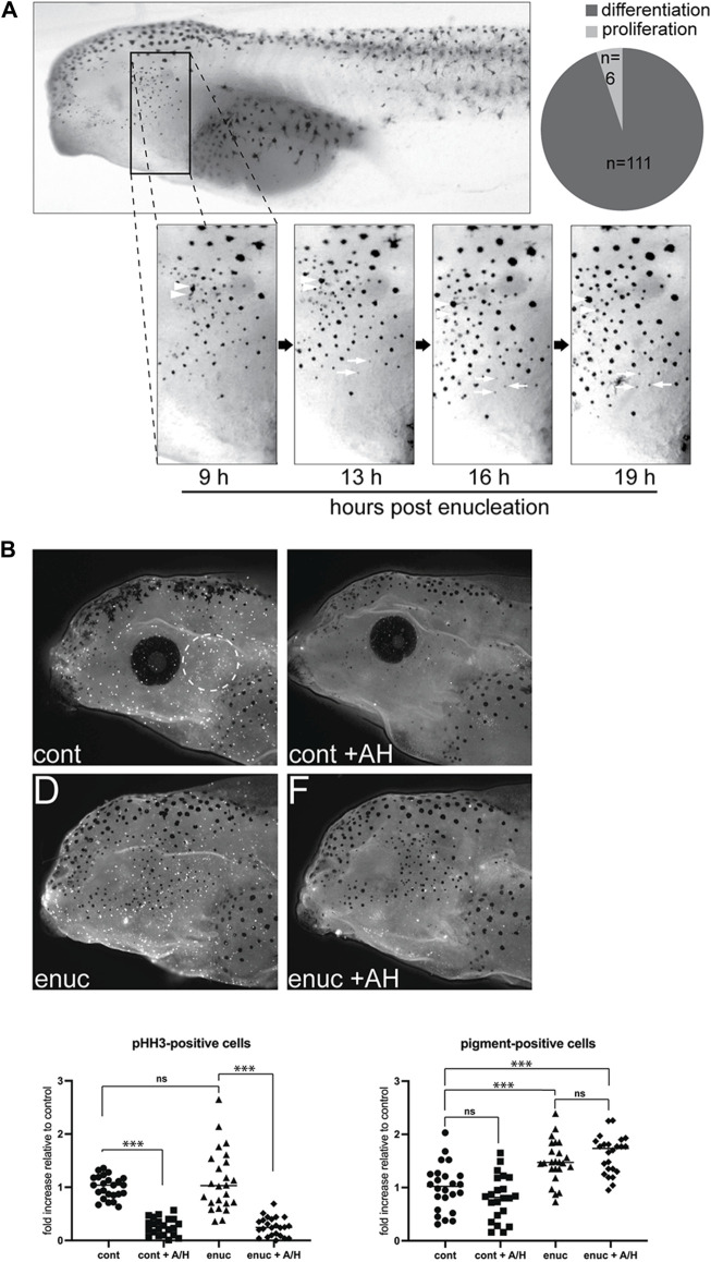
New pigmented cells emerge through de novo pigment production, not migration or proliferation. (A) To track the emergence of new melanophores, enucleated larvae were imaged from 9–19 h post-surgery. A common zone was identified across these images using anatomical landmarks so that individual cells could be tracked over time. Newly emerged pigment cells within the zone were identified (n = 117 melanophores, N = 6). The majority of these (n = 111) appeared de novo as a faint grey dot that darkened over time (white arrows). A few melanophores (n = 6) may have emerged from pigmented cell division (arrowheads). (B) Cell proliferation was inhibited in stage 40 control and enucleated larvae using aphidicolin-hydroxyurea (AH). The numbers of pHH3-positive and pigmented cells were assessed in the perioptic area (white dashed circle) in a blinded fashion after 24 h of AH on a white background. pHH3+ cells in the perioptic region were dramatically reduced in AH-treated larvae (pHH3-positive cells: cont = 1.00 ± 0.04, n cont = 24; cont+AH = 0.26 ± 0.03, n cont+AH = 22; enuc = 1.12 ± 0.12, n enuc = 24; enuc+AH = 0.26 ± 0.04, n enuc+AH = 24; N = 3; F 3,90 = 45.77, p < 0.0001, one-way ANOVA). Whereas the enucleation-induced increase in melanophore number was not impacted (pigment-positive cells: cont = 1.00 ± 0.08; cont+AH = 0.81 ± 0.09; enuc = 1.49 ± 0.08; enuc+AH = 1.62 ± 0.07; F 3,90 = 21.23, p < 0.0001, one-way ANOVA). ***p < 0.0001, Tukey’s test.

