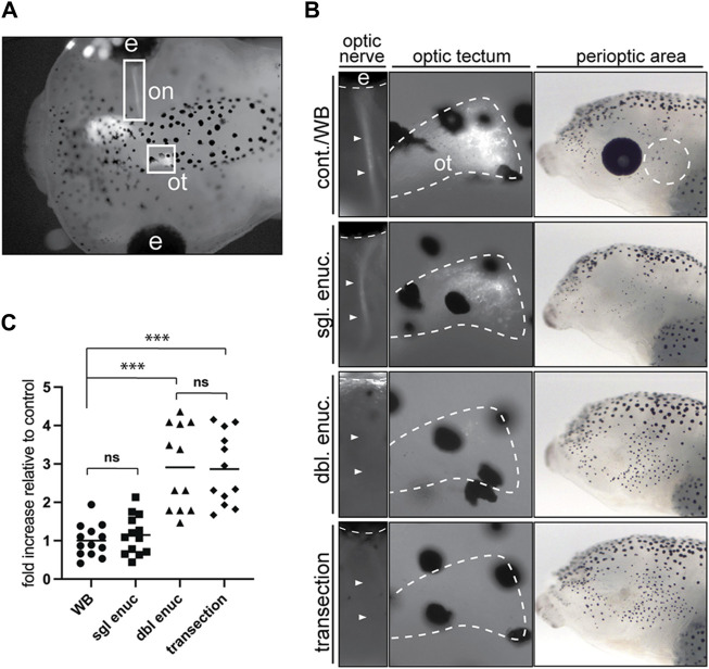
FIGURE 6.
Optic nerve transection mimics the enucleation-induced increase in melanophore number. (A) Larvae with a unilaterally GFP-labeled optic nerve (on) and optic tectum (ot) underwent sham surgery (cont./WB), single enucleation (sgl. enuc.), double enucleation (dbl. enuc.), or single enucleation with transection of the remaining optic nerve (transection). (B) Images of representative larvae from each surgical condition. The first panel shows presence or absence of GFP-positive optic nerve (arrowheads), second panel shows the presence or absence of GFP-positive retinal ganglion cell axon terminals in the optic tectum (white dashed line), and third panel shows lateral brightfield view of perioptic region for each condition. (C) 24 h after enucleation and/or optic nerve transection or sham surgery, melanophore numbers in the perioptic region (white dashed circle in B, third panel) were compared; cont = 1.00 ± 0.12, n cont = 13; sgl enuc = 1.15 ± 0.14, n sgl enuc = 12; dbl enuc = 2.91 ± 0.32; n dbl enuc = 12; trans = 2.87 ± 0.94, n trans = 12; N = 2; F 3,46 = 23.06, p < 0.0001, one-way ANOVA). ***p < 0.0001, n. s., not significant, Tukey’s test.

