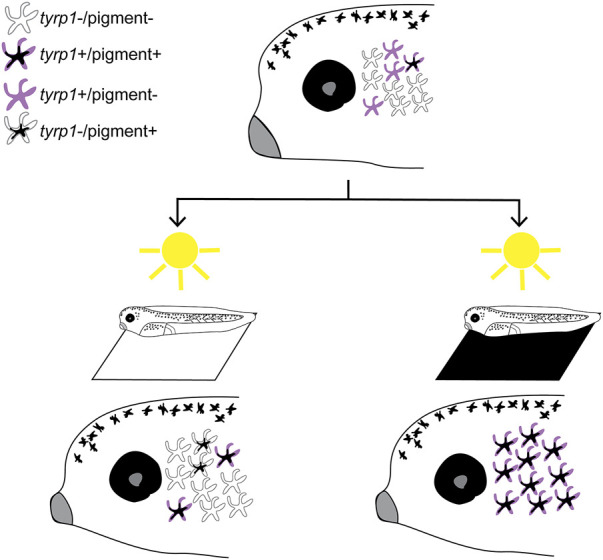
FIGURE 8.

Model of vision-mediated melanophore differentiation. At stage 40 (top), as the visual system becomes functional, four related perioptic melanophores exist: 1) differentiated pigment+/tyrp1+, 2) differentiating tyrp1+/pigment-, 3) immature tyrp1-/pigment-awaiting a signal to differentiate, and 4) de-differentiating tyrp1-/pigment+. Light on a white background (bottom left) produces a small increase in pigmented cells, but tyrp1+ cells (pigmented and non-pigmented) decrease. Light on a black background (or 24 h enucleation; bottom right) dramatically increases pigmented and tyrp1+ melanophores. A black background drives differentiation of the tyrp1-/pigment-population into mature pigmented melanophores.
