Figure 1.
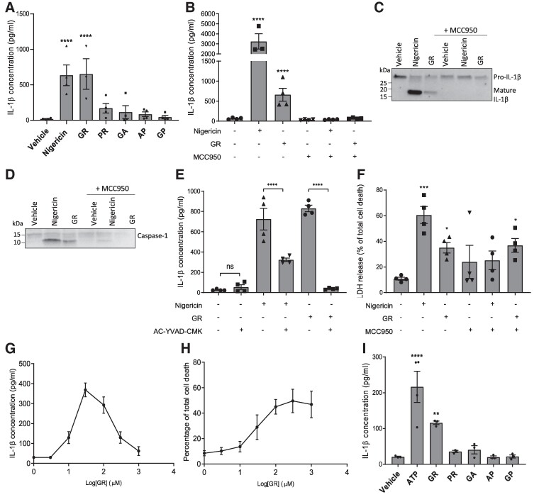
GR activates the NLRP3 inflammasome in macrophages and microglia. (A) LPS-primed primary peritoneal macrophages from male and female WT mice were treated with 30 μM DPRs or controls for 24 h (n = 4). IL-1β was measured in the media by ELISA. (B) GR increased IL-1β release in LPS-primed BMDMs (n = 4) and was prevented by MCC950. (C) Western blot of culture media from GR-treated bone marrow-derived macrophages (BMDMs) showing pro-IL-1β cleavage to produce an active fragment (∼17 kDa). Nigericin was used as a positive control (n = 3). (D) Western blot of culture media from GR-treated bone marrow-derived macrophages (BMDMs) showing pro-caspase-1 cleavage to produce an active fragment (∼10 kDa). Nigericin was used as a positive control (n = 3). (E) GR-induced IL-1β secretion from LPS-primed BMDMs was prevented by AC-YVAD-CMK pre-treatment (n = 4). (F) Quantification of cell death in GR-treated BMDMs by LDH assay, 24 h post-treatment. (G) Dose–response curve of IL-1β detected in BMDM culture media 24 h after treatment with increasing concentrations of GR. (H) Dose–response curve of BMDM cell death measured by LDH assay, 24 h after treatment with increasing concentrations of GR. (I) IL-1β levels in culture media of LPS-primed primary WT mouse microglia 24 h post-treatment with 30 μM DPRs or 5 mM ATP (n = 3). All data analysed by one-way ANOVA with post hoc Dunnett’s or Tukey’s tests for multiple comparisons. **** indicates P < 0.0001, *** indicates P < 0.001, and ** indicates P 0.01. All values are mean ± SEM. Individual mice were considered the experimental unit. See Supplementary Fig. 1 for uncropped blots.

