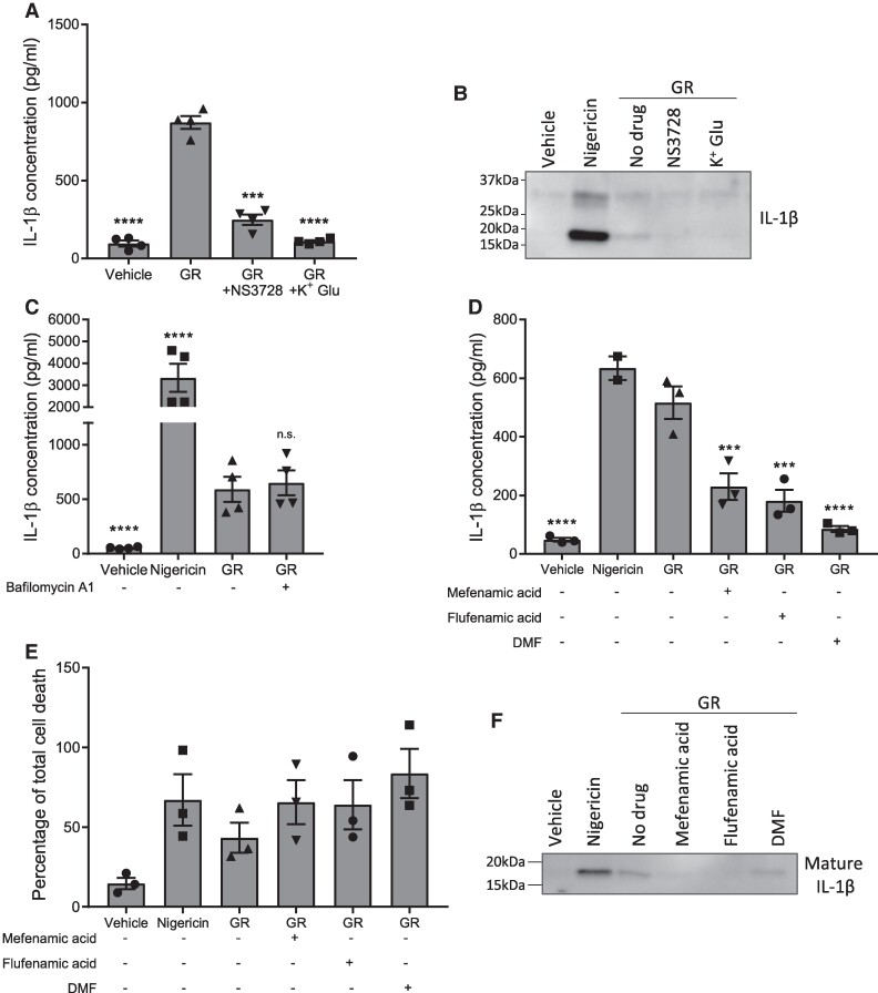
Figure 3.
Characterization of GR-induced NLRP3 inflammasome activation. (A), (B) LPS-primed primary mouse BMDMs (from male and female mice) were pre-treated for 15 min with a chloride channel inhibitor, NS3728, or potassium gluconate (K+ Glu) prior to a 24-h treatment with GR. IL-1β concentrations in the culture media were measured by ELISA (n = 4) (A) and cleavage of pro-IL-1β assessed by WB of the media (n = 3) (B). C LPS-primed BMDMs were pre-treated for 15 min with bafilomycin A1 or vehicle prior to a 24-h treatment with GR, and IL-1β secretion quantified by ELISA (n = 4). (D)–(F) LPS-primed mouse primary BMDMs were pre-treated for 15 min with one of three clinically approved drugs which are known NLRP3 inhibitors: mefenamic acid, flufenamic acid, and dimethyl fumarate (DMF), before a 24-h treatment with GR. (D) Mean IL-1β concentrations in the culture media as detected by ELISA (n = 3). (E) Quantification of cell death in BMDMs assessed by LDH assay (n = 3). (F) Western blotting of the culture media showing cleavage of pro-IL-1β to produce mature IL-1β (17 kDa) (n = 3). All data were analysed by one-way ANOVA with post hoc Dunnett’s or Tukey’s tests for multiple comparisons. The error bars indicate SEM. Individual mice were considered the experimental unit. See Supplementary Fig. 2 for uncropped blots.

