Figure 3.
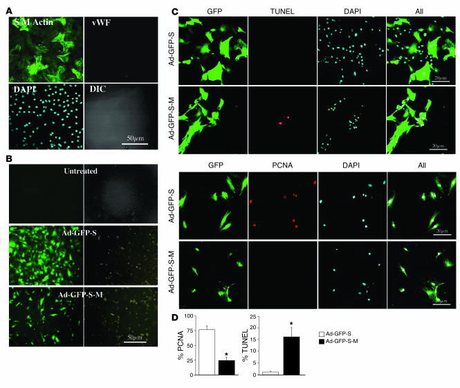
Efficient adenoviral delivery of phosphorylation-deficient survivin (Ad-GFP-S-M) to PASMCs in vitro reduces proliferation and increases apoptosis. (A) Primary culture of rat PASMCs stains positive for smooth muscle actin but not vWF, indicating no contamination with endothelial cells. (B) Infection of PASMCs with adenoviruses encoding GFP and either WT survivin (Ad-GFP-S) or survivin mutant (Ad-GFP-S-M) was highly efficient, as evidenced by GFP reporter (green fluorescence in the left panels and differential interference contrast [DIC] in the right panels). Note the reduced cellularity of the plate infected with Ad-GFP-S-M, compared with Ad-GFP-S. (C) Cells infected with Ad-GFP-S-M (grown in 10% FBS) show increased TUNEL-positive nuclei and reduced PCNA-positive nuclei, suggesting that they undergo apoptosis and not proliferation. In contrast, cells infected with Ad-GFP-S (grown in 0.1% FBS) show no apoptosis and increased rates of PCNA expression. (D) Mean data for TUNEL- and PCNA-positive nuclei, 48 hours after infection with Ad-GFP-S-M versus Ad-GFP-S; 5 fields studied in each plate, 20 plates per group. *P < 0.01.

