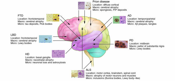
Figure 3.
Overview of the anatomical location of and macroscopic and microscopic changes characteristic of the neurodegenerative disorders discussed in this review. Note that the full neuropathological spectrum of these disorders is much more complex than depicted here. When there is more than one characteristic histopathological feature, these are depicted from left to right, as indicated in the labels listing microscopic changes (e.g., the 2 panels for AD depict an Aβ plaque [left] and neurofibrillary tangles [right]). All histopathological images are reprinted with permission from ISN Neuropath Press (ref. 99).

