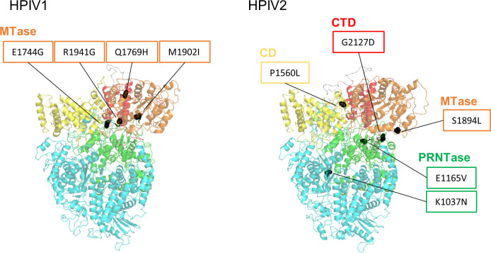
Fig 4.
Amino acid mutations occurring in L protein. The cartoon representation shows the position of amino acid residues (black sphere), highlighting the mutations >50% variant frequency shown in Table 3. The domains are described based on previous reports (19): RNA-dependent RNA polymerase (RdRp), cyan; poly-ribonucleotidyltransferase (PRNTase), green; connecting domain (CD), yellow; methyltransferase (MTase), orange; C-terminal domain (CTD), red.

