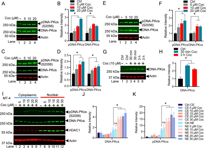
Figure 1: Cocaine enhances both the catalytic activity and nuclear translocation of DNA-PK.
Jurkat cells harboring the pHR’-P-Luc provirus (A), microglial cells (C), and MT-4 cells (E) were treated with different concentrations of cocaine (Coc: 5, 10, and 20 μM) for 3 h (Lanes 2 to 4). Jurkat-pHR’-P’-Luc cells were treated with 10 µM cocaine (Coc) in replicates for 30 min and 3 h (Lanes 3 to 6) (G). Cells were harvested, and nuclear lysates were analyzed by immunoblotting using specific antibodies, pDNA-PKcs (S2056) and DNA-PKcs, as indicated. Actin, a constitutively expressed protein, was used as a loading control. Densitometric analysis of protein bands (normalized to actin) confirmed the significant upregulation of total DNA-PKcs and its phosphorylated form, pDNA-PKcs S2056 (pDNA-PKcs), following cocaine treatment (B, D, F, & H). MT-4 cells were treated with increasing doses of cocaine for 3 h. Cells were harvested and lysed, and both cellular and nuclear lysates were analyzed by immunoblotting with antibodies against pDNA-PKcs (S2056), DNA-PKcs, HDAC1, and Actin (I). Densitometric analysis of protein bands, normalized to actin, validated the enhancement in both the catalytic activity and nuclear translocation of DNA-PK (J & K). Immunoblots are representative of at least three independent experiments. The results are expressed as mean ± SD and analyzed by one- or two-way ANOVA, followed by Tukey’s multiple comparison test. Asterisks over the bars indicate significant differences: ∗p < 0.05 for the comparison of cocaine-treated cells vs. untreated cells (Ctrl).

