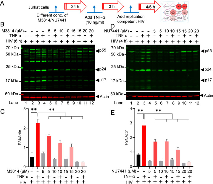
Figure 4: Partial DNA-PK inhibition restricts HIV replication.
Schematic timeline for the treatment with M3814, NU7441 inhibitors, TNF-α, and replication-competent HIV (A). Jurkat cells were treated overnight with different concentrations of M3814 (5, 10, 15, and 20 μM) (B) and NU7441 (5, 10, 15, and 20 µM) (D) for 24 h (Lanes 5–12). The next day, cells were activated with 10 ng/ml TNF-α for 3 h (Lanes 3, 4, 6, 8, 10, & 12). Subsequently, cells were infected with a replication-competent dual-tropic HIV (Type 1 strain 93/TH/051) (Lanes 1–12). Cell lysates were prepared 4 h (NU7441) or 6 h (M3814) post-infection (hpi). Total cell lysates were analyzed by SDS-PAGE, transferred to a nitrocellulose membrane, and detected with specific HIV antibodies as indicated. Immunoreactive proteins were detected using appropriately labeled secondary antibodies with Licor. Actin was used as a loading control. Densitometric analysis of protein bands relative to actin (C & E). Immunoblots are representative of at least three independent experiments. The results are expressed as mean ± SD and analyzed by one-way ANOVA followed by Tukey’s multiple comparison test. Asterisks over the bars indicate significant differences: **p < 0.01 for the comparison of inactive vs. activated cells (TNF-α) and activated cells (TNF-α) vs. activated cells (TNF-α) in the presence of DNA-PK inhibitors, NU7441 or M3814.

