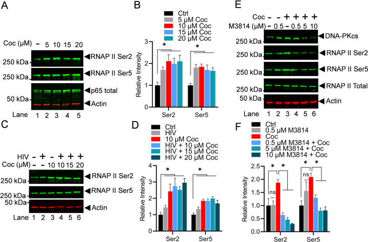
Figure 6: Cocaine promotes HIV transcription by enhancing the phosphorylation of the C-terminal domain (CTD) of RNA polymerase II (RNAP II).
THP-1 cells were treated with increasing doses of cocaine (5, 10, 15, and 20 µM) for 3 h (A). MT-4 cells were treated as follows: untreated and uninfected (Lane 1), infected with HIV (93/TH/051) without cocaine treatment (Lane 2), treated with cocaine without HIV infection (Lane 3), or pre-treated with different concentrations of cocaine before HIV infection (Lanes 4 to 6) (C). Cells were harvested, and nuclear lysates were analyzed by immunoblotting with specific antibodies against phosphorylated RNAP II, RNAP II Ser2, and RNAP II Ser5. Actin, a constitutively expressed protein, was used as a loading control. Densitometric analysis of protein bands (normalized to actin) confirmed significant hyper-phosphorylation of RNAP II CTD at both Ser2 and Ser5 residues following cocaine treatment (B & D). THP-1 cells were treated with cocaine in the absence or presence of different concentration of M3814 (0.5, 5, and 10 µM) (E). Cells were harvested, and nuclear extracts were evaluated via immunoblotting using specific antibodies against RNAP II Ser2, RNAP II Ser5, and total RNAP II. Densitometric analysis of protein bands (normalized to actin) confirmed a significant increase in RNAP II CTD phosphorylation at both Ser2 and Ser5 upon cocaine treatment. However, a significant reduction in CTD phosphorylation at both Ser2 and Ser5 was observed upon DNA-PK inhibition with M3814 compared to cocaine-alone samples (F). Immunoblots are representative of at least three independent experiments. The results are expressed as mean ± SD and analyzed by two-way ANOVA followed by Tukey’s multiple comparison test. Asterisks over the bars indicate significant differences. ∗ p < 0.05 is for the comparison of cocaine-treated samples against untreated (Ctrl) and the comparison of cocaine plus inhibitors treated against cocaine alone-treated samples

