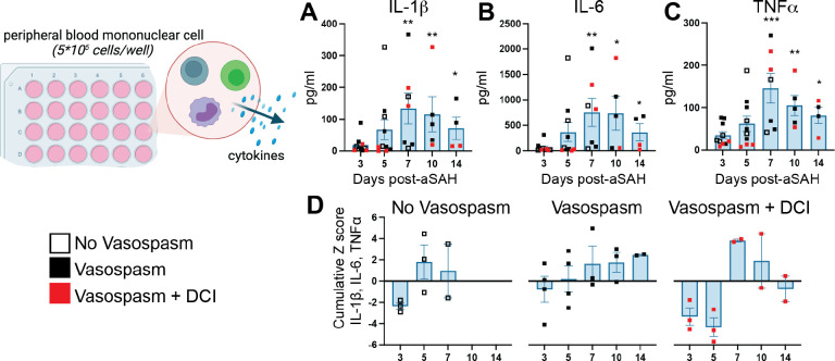
Figure 3: Basal cytokine production by PBMCs increases post-aSAH.
Supernatants from unstimulated PBMCs incubated for 40 hours were measured 3–14 days post-aSAH. (A) IL-1β peaked around day 7 (p=0.0220). (B) IL-6 was significantly higher at day 7 (p=0.0236). (C) TNFα showed significant increases on days 7 (p=0.0149) and 10 (p=0.0452) compared to day 3. (D) A pro-inflammatory cytokine cumulative z-score of IL-1β, IL-6, and TNFα, stratified by vasospasm and DCI status, revealed a unique profile of proinflammatory changes. The Vasospasm+DCI group failed to activate a proinflammatory response to the injury at days 3–5 but showed a reversal at day 7. Bar graphs plot mean values; error bars indicate SD. The main effects were analyzed using a one-way repeated measures ANOVA (mixed-effects model REML). Student’s t-tests were used to compare day 3 vs. day 5, day 3 vs. day 7, and day 3 vs. day 10. *P<0.05, **P<0.01, ***P<0.001. Data is from 10 patients. See also Table S3 for all analytes measured.

