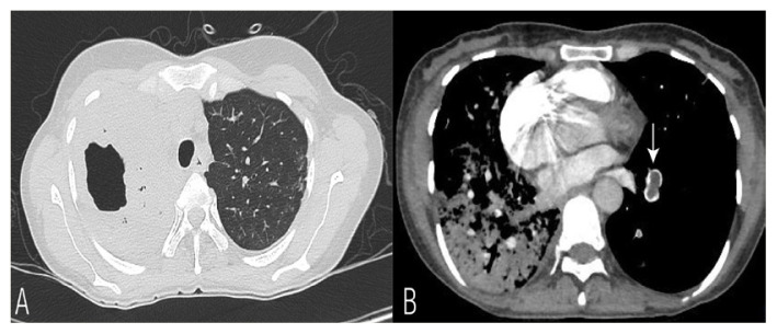
Figure 2.
A: Parenchymal window of computed tomography (CT)thorax showing dense consolidation in right hemithorax with a cavity in right upper lobe. B: CT pulmonary angiogram image revealing hypodense thrombus in the arterial branch of left lower lobe (vertical arrow) highly suggestive of pulmonary thromboembolism.

