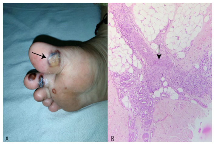
Figure 3.
A: Photograph showing haemorrhagic vesicles with perilesional purpura and erythema over the sole region (arrow). B: Haematoxylin and eosin stain of subcutaneous tissue at ×100 magnification showing medium-sized vessels infiltrated by histiocytes in aggregates, lymphocytes and a few neutrophils. The vessel wall shows focal fibrinoid necrosis (arrow).

