
FIG 3.
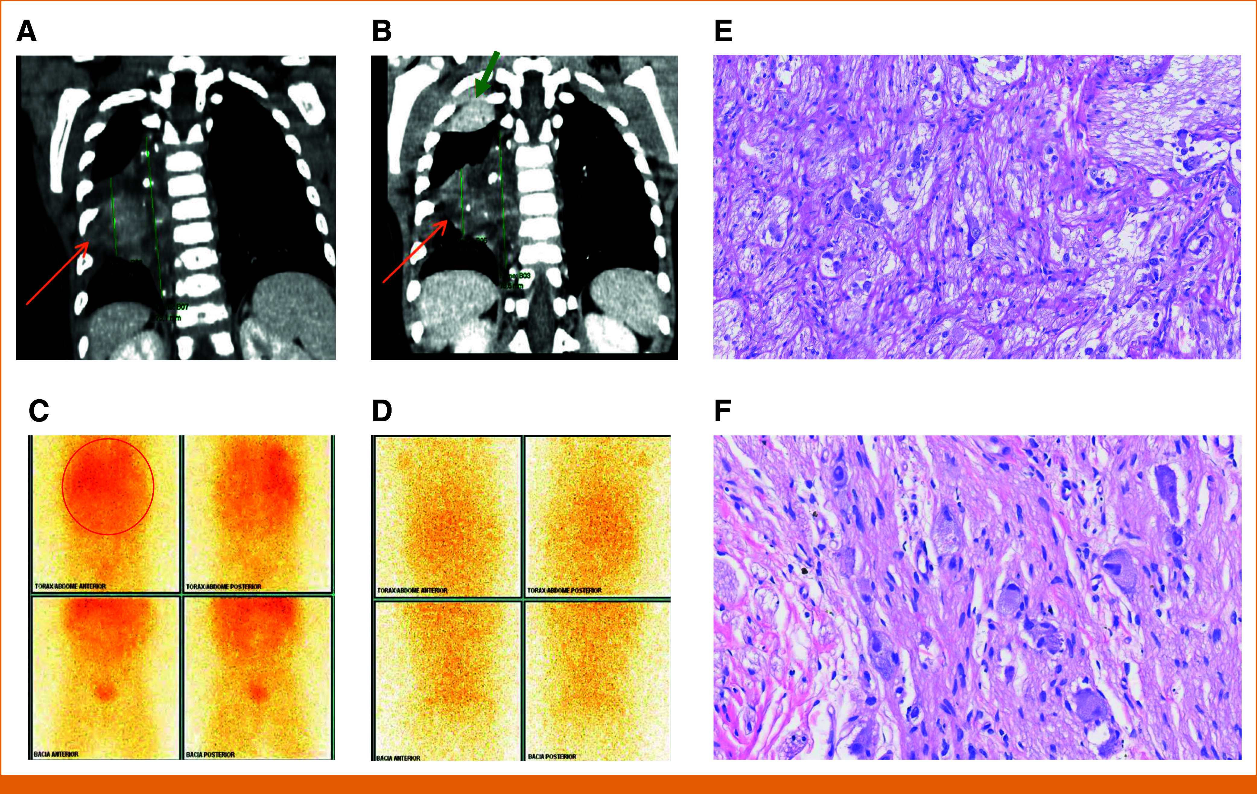
The chest CT scan (soft tissue window, coronal reformatting) shows a heterogeneous right paravertebral mass in the mid thoracic segment, with (A) hypoattenuating areas and avid uptake on the (C) PET-MIBG study. (B) After treatment, there was a slight reduction in size (being characterized as a stable disease by RECIST) and the onset of small foci of calcification (orange arrows) and (D) no more PET uptake. (B) Also, an atelectatic opacity was observed at the apex of the right lung, probably related to decubitus (green arrow). The histopathological study of specimens before and after larotrectinib treatment shows (E) a poorly differentiated NB specimen and (F) a completely mature NB tissue compatible with ganglioneuroma. CT, computed tomography; MIBG, metaiodobenzylguanidine; NB, XXX; PET, positron emission tomography.