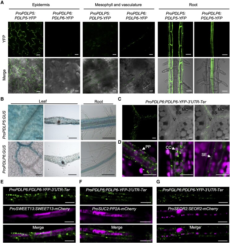
Figure 3.
PDLP5 and PDLP6 are expressed in different cell types. A) The cell type–specific expression of PDLP5-YFP and PDLP6-YFP. The fusion proteins were driven by their own native promoter. Confocal images were captured from 2-wk-old Arabidopsis seedlings. The signal of the YFP fusion proteins was detected in different cell types (top panel). Merged images show the signals from YFP and bright-field (lower panel). Scale bars for epidermis and vasculature = 10 µm. Scale bars for root = 50 µm. B) Histochemical staining of GUS activity in Arabidopsis ProPDLP5:GUS and ProPDLP6:GUS transgenic plants. Images were captured from the leaf surface, leaf transverse sections, and roots. Scale bars = 100 µm. C) PDLP6-YFP proteins were observed in the vasculature. The leaf sample was cleared by ClearSee solution. MC, mesophyll cell; BS, bundle sheath cell; V, vasculature. Scale bars = 10 µm. D) PDLP6-YFP proteins were detected in PP cells, CC, and SE. Cell types were determined based on their sizes, chloroplast arrangement, and cell wall ingrowth phenotype as previously described (Cayla et al. 2015). Chlorophyll autofluorescence was also shown. Scale bar = 10 µm. E to G) The colocalization of PDLP-YFP signals with various cell type markers. E)ProSWEET13:SWEET13-mCherry marks PP cells. F)ProSUC2:PP2A1-mCherry marks CC. G)ProSEOR2:SEOR2-mCherry makes SE. Scale bars = 10 µm.

