Figure 3.

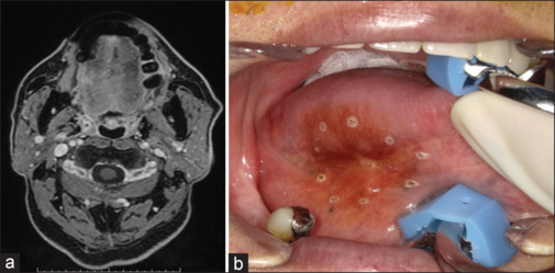
(a) MR image after completion of intra-arterial DCF therapy in case 1. (b) Intraoral photograph at the time of surgery. The excisional margin was inside the marking points

(a) MR image after completion of intra-arterial DCF therapy in case 1. (b) Intraoral photograph at the time of surgery. The excisional margin was inside the marking points