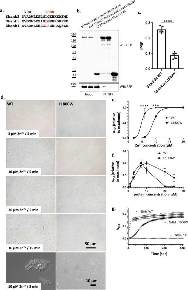
Fig. 2. p.L1800W mutation in the SAM domain interferes with Shank2 homo-oligomerization.
a Alignment of sequences surrounding L1800 (red) in the Shank2-SAM domain with those of other Shank SAM domains. b GFP-tagged Shank2b, or GFP control, was coexpressed with mCherry-tagged variants of Shank2a in 293T cells. After cell lysis, GFP-tagged proteins were immunoprecipitated using the GFP-trap matrix. Input and precipitate samples were analysed with GFP- and mRFP-specific antibodies. c Quantification of the data shown in (b). ****, significantly different, p < 0.0001; data from five independent experiments; t test. d His6-SUMO tagged Shank SAM domains (WT and L1800W mutant) at a concentration of 10 µM were treated with various concentrations of Zn2+. After 5 min (or 15 min, lower panel), 20 µl drops were spotted on a microscopic slide and photographed through a phase contrast microscope. Higher resolution DIC images were also generated (lower panels). Bars: 50 µm, 10 µm. e Samples containing 10 µM SAM domain were treated with different concentrations of Zn2+ in wells of a 96-well microtiter plate. Absorbance at 350 nm was measured after 10 min. Data were normalized to the maximum absorbance obtained in each experiment, and analyzed by nonlinear regression using GraphPad Prism software. ****, ***: significantly different, p < 0.0001, 0.001, respectively; t test. f Measurements were performed as in (d), however the Zn2+ concentration was kept constant while protein concentration was varied as indicated. g Protein samples at a concentration of 10 µM were treated with 10 µM Zn2+. After the start of the reaction, absorbance was measured at 350 nm every 10 s. Here a His6-SUMO fusion protein containing SH3 and PDZ domains of Shank2 was included as a negative control. In (e–g), mean ± SD of 4 independent experiments is shown.

