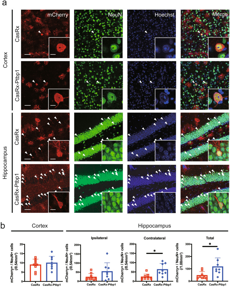
Fig. 3.
(a) Immunofluorescent staining of mCherry/NeuN/Hoechst in the cortex and hippocampus 56 days after tMCAO. Arrowheads indicate mCherry/NeuN double-positive cells. Scale bars: 50 µm and 10 µm. (b) Quantitative analysis of mCherry and NeuN double-positive cells in the cortex and hippocampus. CasRx-Ptbp1 significantly increased the number of mCherry/NeuN double-positive cells in the DG compared with the CasRx group (*p < 0.05). P-values < 0.05 were considered statistically significant.

